
We integrate human iPSC disease models, functional phenotyping, molecular profiling, and proprietary gene network analysis to reveal disease mechanisms, prioritize therapeutic targets, and support confident discovery decisions.
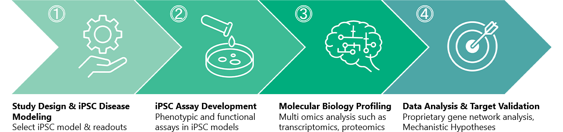
We combine human iPSC disease models, functional and molecular readouts, and proprietary gene network analysis to connect an observed phenotype to the biology producing it, and that mechanism to a prioritized set of therapeutic targets.
Human iPSC-derived neurons, astrocytes, and microglia in advanced mono-, co-, and tri-culture models, enhanced with cellular aging technologies to better recapitulate late-onset neurodegenerative diseases.
Functional and molecular readouts chosen so an observed phenotype can be traced back to the biology behind it, rather than recorded on its own.
Network-level analysis over RNA-Seq and microarray data, turning molecular output into mechanistic hypotheses and candidate targets. See what the analysis returns.
Fujifilm began drug discovery research for central nervous system (CNS) disorders following the acquisition of Toyama Chemical Co., Ltd. in 2008, developing drug evaluation systems based on human iPSC-derived neural cells alongside gene network analysis techniques. The 2015 acquisition of Cellular Dynamics International further deepened our iPSC-derived cell expertise. Since 2023 we have offered these capabilities as discovery support services, with particular depth in CNS disorders.
Proprietary cellular aging and tri-culture technologies let us tailor high-throughput pharmacological assays to your program.
- Alzheimer's disease
- tau biology, neurotoxicity, neuroinflammation
- Parkinson's disease
- dopaminergic neuron dysfunction
- Amyotrophic lateral sclerosis (ALS)
- oxidative stress, TDP-43 pathology.
- Neuroinflammation
- neuron, astrocyte and microglia interaction models
- Neurodegeneration
- neuronal survival and neurite integrity
- Pain
- sensory neuron models
- iPSC-derived neurons | GABA, Gluta, Dopa and Motor
- iPSC-derived microglia
- iPSC-derived astrocytes, young and original aged
- Patient-derived models
- Mutation-engineered models
- 2D and 3D mono-, co- and tri-culture
- Neuroinflammation in tri-culture
- Oxidative stress-induced neuronal damage
- Neurotoxic astrocyte model
- Blood-brain barrier (BBB) models
- ASO Assessment Platform: target engagement, efficacy and inflammatory-liability evaluation
Mono-culture: Identify direct effects on specific cell types.
Co-culture: Reveal cell-cell interactions and indirect effects.
Tri-culture: Understand complex neuroinflammatory mechanisms and compound responses.
- Neurite length
- Oxidative stress-induced damage
- TDP-43 aggregation model
- Aβ / tau / p-tau ELISA
- Reactive-state markers
- Age-associated studies
- Neurotoxic-associated phenotype
- Gene knockdown
- Phagocytosis assay
- Cytokine response
- Viability and metabolic activity
- Molecular profiling
At Fujifilm Drug Discovery Services, we specialize in establishing innovative translational in vitro disease models, focusing on Alzheimer's Disease and Parkinson's Disease, among others. Our ALS model employs chronic oxidative stress-mediated neuronal damage in iPSC-derived iCell® Motor Neurons and links clinical results with targeting ferroptosis and cholesterol biosynthesis, both of which are increasingly recognized for their involvement in ALS development.
Our method of introducing antioxidant deficiency leads to oxidative stress in iCell® Motor Neurons, which results in neurodegeneration. This process is mitigated by Edaravone, a clinically approved ALS therapeutic drug.


Ferroptosis inhibitors effectively prevented neuronal damage caused by gradual oxidative stress, whereas inhibitors of apoptosis or necroptosis did not exhibit similar neuroprotective benefits.
Apoptosis inhibition
Necroptosis inhibition
Ferroptosis inhibition
Distal cholesterol biosynthesis inhibitor AY 9944 inhibits neurodegeneration in the chronic oxidative stress model, possibly by leading to an accumulation of 7-DHC, which is a precursor of cholesterol.
Compound screen


Cholesterol Biosynthesis
Neuronal Damage Induced by Gradual Oxidative Stress in iPSC-Derived Neurons: Implications for Ferroptosis Involvement and ALS Drug Evaluation (Journal of Neurochemistry, Vol.169, 2025, e70246)
Hayato Kobayashi, Hitoshi Suzuki-Masuyama, Hirokazu Tanabe, Hiroshi Kato, Setsu Endoh-Yamagami
How do two interacting cell types influence one another?
- Astrocyte neurotoxicity assay
- Calcium imaging
- MEA
- Neurite and network integrity
- Microglial effects on neurons
- Inflammatory signaling
- Cytokine analysis
- Neuronal viability and network effects
- Morphology assay
- Cytokine ELISA
iPSC reprogramming resets age-associated cellular features, which is a real mismatch when the disease being modeled is late-onset. Prolonged culture without chemical induction produces cryo-preservable astrocytes carrying multiple age-associated features, so age becomes a variable you choose rather than one you inherit.
TNF-α and IFN-γ induce neurotoxicity in human iPSC-derived astrocytes that is enhanced by aging, (Scientific Reports,Published: 05 September 2026)
We utilize human iPSC-derived neuronal and glial cells for CNS drug discovery and development, focusing on neurodegeneration and neuroinflammation, among other models. Our proprietary cell aging and co-culture technologies enable customized HTS pharmacological assay services tailored to your needs.
Advanced model for detection of neuroprotective compounds utilizing iPSC-neurons and aged iPSC-astrocytes.

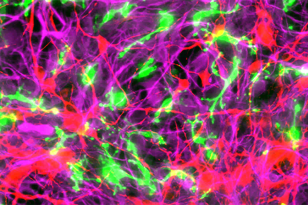
iCell neuronal cell types

iCell neuronal cell types
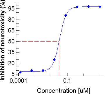
Inhibition of astrocyte-mediated neurotoxicity
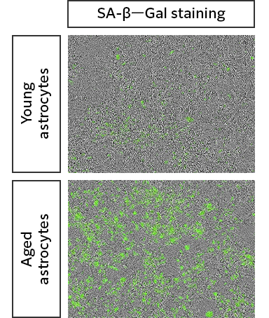
Against young astrocytes, the prolonged-culture cells show multiple senescence-associated features at once, increased γH2AX signal, increased SA-β-Gal activity and increased CDKN2A mRNA, rather than a single marker moving on its own. Supporting preprint: bioRxiv 2025.08.17.670762.
How does inflammation propagate across the network?
- 2D tri-culture (neuron / astrocyte / microglia)
- 3D brain-like culture, where applicable
- Neuroinflammation assay
- Cell-type specific NF-κB activation
- Cytokine expression and secretion
- Neuronal calcium activity
- Neuronal network integrity
- Mechanism-focused pathway perturbation
In tri-culture microglia adopt a more branched morphology than in monoculture, closer to their state in tissue, and the inflammatory response to LPS is amplified relative to co-culture. The added cell types in cell culture changes both the baseline phenotype and the magnitude of the response.
with Complex Tri-culture Systems of iPSC-Derived Microglia, Astrocytes, and Neuron
- Optional study-specific profiling can be added to any format: staining and imaging analysis, cytokine assays, Ca2+ imaging, MEA, gene expression analysis, EV isolation and analysis, gene network analysis and omics. '- Functional Neuronal Assays: Characterize neuronal activity using calcium imaging and multi-electrode array analysis.
- Functional Neuronal Assays: Characterize neuronal activity using calcium imaging and multi-electrode array analysis.
- ASO Assessment Platform: Evaluate target engagement, efficacy, and inflammatory liabilities.
Using proprietary algorithms, we mine gene expression data from RNA-Seq and microarray studies to visualize upstream and downstream relationships between genes, letting researchers hypothesize drug mechanisms and identify key gene targets.
The core strength is the combination of advanced variable compression techniques, high-efficiency supercomputing resources, and the joint expertise of pharmacologists and computational biologists, surfacing connections between genes that conventional methods often miss.
Beyond interpretability, the analysis gives actionable insight into processes such as aging, inflammation and disease progression, identifying candidate therapeutic targets through network analysis.
- Pathways associated with disease progression
- Candidate central regulators within those pathways
- Hypotheses about the mechanisms driving the disease
- A prioritized set of candidate therapeutic targets

Inferred gene network showing upstream and downstream relationships.

A worked result: clusters read as decreased astrocyte differentiation and function alongside an increased inflammation signal response.
The core strength is the combination of advanced variable compression techniques, high-efficiency supercomputing resources, and the joint expertise of pharmacologists and computational biologists, surfacing connections between genes that conventional methods often miss.
Beyond interpretability, the analysis gives actionable insight into processes such as aging, inflammation and disease progression, identifying candidate therapeutic targets through network analysis.
Best deployed within a comprehensive CNS discovery package to maximize biological insight and translational relevance. Supporting publication: Maeda K, “Gene grouping strategy for network modeling from a small time-series dataset”, Biosystems 179 (2019) 24–29.
From phenotype to molecular insight: functional, cellular and molecular readouts, combined to show how targets and compounds affect disease-relevant biology.
- Define the Biological Question
Examine the existing evidence to establish the question the study needs to answer. - Build and Characterize the Disease Model
Build the disease model and culture format appropriate to your target. In a Parkinson's disease program, for example, this means a monoculture hiPSC-derived neural cell model. - Generate Functional and Molecular Data
Execution of the agreed pharmacological, phenotypic and molecular readouts — for a Parkinson's program, neuronal survival and neurite length as the functional readouts, paired with an RNA-seq dataset as the molecular one. - Integrate Data and Develop Mechanistic Hypotheses
Pathway analysis over the assay readouts, drawing on gene network analysis when a hypothesis needs confirming or an upstream regulator needs finding. - Gene network analysis [optional]
Available as part of a study or as a standalone service. See what the analysis returns. - Prioritize and Validate Therapeutic Targets
Confirmation by gene knockdown, producing disease drivers, a mechanism hypothesis and target candidates as deliverables.
- PD Disease Modeling using hiPSC derived neural cells
- Phenotypic and Functional readouts
Neuronal survival
Neurite length - Molecular Biological Profiling
RNA-seq dataset - Data Analysis
Gene Network Analysis - Deliverables
Disease drivers
Mechanism hypothesis
Target candidates
We build assay models around your research question, so service deliverables can vary. Connect directly with our scientists to scope a study.
- TNF-α and IFN-γ induce neurotoxicity in human iPSC-derived astrocytes that is enhanced by aging, (Scientific Reports,Published: 05 September 2026) Hayato Kobayashi, Kiyohiro Maeda, Takashi Wakui, Kenichi Kazetani, Akira Nabetani, Mitsuho Taniguchi, Euikyung Shin, Hiroshi Kato & Setsu Endoh-Yamagami
- Tri-Culture System Reveals an Activation Cascade From Microglia Through Astrocytes to Neurons During Neuroinflammation (Journal of Neurochemistry, Volume170, Issue3, March 2026) Hayato Kobayashi, Hiroshi Kato, Mitsuho Taniguchi, Setsu Endoh-Yamagami
- Neuronal Damage Induced by Gradual Oxidative Stress in iPSC-Derived Neurons: Implications for Ferroptosis Involvement and ALS Drug Evaluation (Journal of Neurochemistry, Volume169, Issue10, October 2025) Hayato Kobayashi, Hitoshi Suzuki-Masuyama, Hirokazu Tanabe, Hiroshi Kato, Setsu Endoh-Yamagami
- Induction of neuroinflammation in iPSC-derived neuron-astrocyte-microglia tri-culture systems and their application in ASO toxicity assays (H. Kobayashi, H. Munezane, and S. Endoh-Yamagami)
- PD model using iPSC-derived tri-culture system reveals induction of neurotoxicity by stimulation with α-synuclein and IFN-γ
(H. Kobayashi, S. Endoh-Yamagami)
- Induction and dynamics of TDP-43 aggregation in iPSC-derived neurons (Setsu Endoh-Yamagami, Hayato Kobayashi,)
- Parkinson's disease (PD) model composed of an iPSC-derived tri-culture system of neurons, astrocytes, and microglia stimulated with α-synuclein (Hayato Kobayashi, Setsu Endoh-Yamagami)
- Tau aggregation induces cell death in iPSC-derived neurons (Aging Brain Volume 7, 2025, 100136)
Hirokazu Tanabe, Sumihiro Maeda, Etsuko Sano, Norio Sakai, Setsu EndohYamagami, Hideyuki Okano - MAPT-A152T mutation drives neuronal hyperactivity through Fyn-NMDAR signaling in human iPSC-Derived neurons: Insights into Alzheimer's pathogenes (Regenerative Therapy Volume 28, March 2025, Pages 201-213)
Maika Itsuno, Hirokazu Tanabe, Etsuko Sano, Takashi Sasaki, Chisato Oyama, Hiroko Bannai, Koichi Saito, Kazuhiko Nakata, Setsu Endoh-Yamagami, Hideyuki Okano, Sumihiro Maeda - Neuronal Damage Induced by Gradual Oxidative Stress in iPSC-Derived Neurons: Implications for Ferroptosis Involvement and ALS Drug Evaluation (Journal of Neurochemistry, Vol.169, 2025, e70246)
Hayato Kobayashi, Hitoshi Suzuki-Masuyama, Hirokazu Tanabe, Hiroshi Kato, Setsu Endoh-Yamagami
- Ferroptosis-dependent neuronal damage induced by oxidative stress and TDP-43 aggregation formation in iPSC-motor neurons as ALS models (Hayato Kobayashi, Hitoshi Suzuki-Masuyama, Hirokazu Tanabe, Hiroshi Kato, and Setsu Endoh-Yamagami)
- iPSC-derived senescent astrocytes generate inflammation prone environment and they are more sensitive to cytokines inducing a neurotoxic state (H. Kobayashi, T. Wakui, H. Kato, S. Endoh-Yamagami)
- Ferroptosis-dependent neuronal injury induced by oxidative stress in iPSC-derived motor neurons as ALS model (H. Kobayashi, H. Suzuki-Masuyama, H. Tanabe, H. Kato, S. Endoh-Yamagami)
- Study of glial function on neuronal activity under neuroinflammation in neuron/astrocyte/microglia tri-culture system (H. Kobayashi, H. Kato, S. Endoh-Yamagami)
- Evaluation system for seizure risks using a co-culture system consisting of human iPS cell-derived neurons and astrocytes (M. Taniguchi, H. Kobayashi, S. Endoh-Yamagami)
- Neurotoxic human iPSC-astrocytes and gene expression network analysis to identify drug targets for neurodegenerative diseases (Hayato Kobayashi, Kiyohiro Maeda, Kenichi Kazetani, Akira Nabetani, Setsu Endoh-Yamagami)
- 2D and 3D tri-culture systems for investigation of cellular interaction among neurons, astrocytes, and microglia (Hayato Kobayashi, Setsu Endoh-Yamagami)
- Induction and characterization of human A1 astrocytes (H. Kobayashi, A. Nabetani, H. Suzuki-Masuyama, W. Shin, K. Maeda, K. Kazetani, T. Wakui, J. Hosokawa, S. Endoh-Yamagami)
Our iPSC-based CNS drug discovery services are trusted by leading global pharmaceutical and biotechnology companies to support drug discovery programs.
Explore how human iPSC disease models and data-driven analysis can support your target discovery and validation strategy.